
Abductor tendon problems are a common source of Greater Trochanteric Pain Syndrome (GTPS) that fails to respond to conservative treatment for what is often erroneously described as “recalcitrant trochanteric bursitis”. When symptoms do not respond to initial treatment, an MRI may be necessary to look for signs of tendon damage. When identified, ultrasound-guided injections may be appropriate. The damage can usually be well visualized under ultrasound, and injection of anesthetic is used as a diagnostic test to differentiate that the tendon damage is truly the source of pain. (Related Study: Perioperative Care) Along with this, cortisone can be used for potential therapeutic benefit. If the patient responds well, repeat injection may be appropriate, but multiple injections should be avoided. This just starts to treat the symptoms and not address the problem, which can worsen.
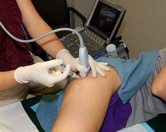
The area of damage can be precisely injected with ultrasound guidance. Relief confirms that the damage is the source of pain; and can sometimes help resolve the problem.
Partial tears that fail conservative treatment can be addressed with endoscopic repair. This includes freshening up the torn part of the tendon and fixing it back to bone. Not all complete tears require surgery as some may be asymptomatic, but complete tears can be repaired endoscopically as well. (Related Studies: Peritrochanteric Access and Gluteus Medius Repair and Gluteus Medius Repair With Double-Row Fixation) There are some circumstances where a large complete tear may be better managed with an open procedure to maximize the security of the repair. Again, many tears do not require surgery at all. But under the correct circumstances, the experience at NHI is that the results of endoscopic repair can be quite gratifying. (Related Study: Endoscopic repair of hip abductor tears: outcomes with two-year follow-up)
Gluteal tears are accessed from the Peritrochanteric Space. A variety of methods are available to repair the tendon depending on the tear pattern.
The goal is to create a repair construct that is as secure as possible. A Double Row method is one way of accomplishing this.
With chronic tears, the tendons may retract, and the muscles may atrophy, with nothing left to repair. However, all is not lost. Even in these circumstances, when the symptoms are disabling, reconstructive methods are available. This involves taking a portion of the gluteus maximus tendon from the back and the tensor fascia lata from the front and bringing them together over the greater trochanter to recreate the abductor group. While this involves an open technique, it can be very effective in the right circumstances.
Rehabilitation following abductor tendon surgery is prolonged. (Related Study: Rehabilitation of the Hip) It is certainly worth it in the face of disabling symptoms, but is a procedure for which the patient needs to be prepared to dedicate themselves to the postoperative rehabilitation process. Crutches or a walker are necessary for 6-8 weeks, with ongoing precautions to protect the repair site for the first 4 months. Typically, it takes about 6 months to really get beyond the surgery and appreciate the benefits.
Location
2004 Hayes Street
Suite 700
Nashville, TN 37203
Office Hours
Monday-Friday:
8:00 am – 5:00 pm